

Understanding Pedal Bone Fractures
- Marc Jerram

- Aug 3, 2025
- 6 min read
Introduction
Pedal bone fractures in horses sometimes referred to as fractures of the coffin bone, are serious injuries that occur inside the hoof. Although the pedal bone (the lowest bone in the horse’s leg) is protected by the hard hoof wall, it can still break due to trauma such as a kick, stepping on a rock, hard landing during exercise, or sometimes because of repeated stress over time. These injuries can cause sudden and severe lameness, and while they are serious, many horses recover well with proper treatment. This article will explain what these fractures are, how they are diagnosed, and the different ways they can be treated using veterinary care, specialist shoeing, and good management practices.
Anatomy
The pedal bone is a triangular-shaped bone that sits inside the hoof capsule. It plays a key role in supporting the horse’s weight and helping absorb the forces of movement. Because it is hidden inside the hoof, damage to this bone isn’t always obvious from the outside. Fractures in this area can happen in many different patterns, depending on how the injury occurred and how much of the bone is affected. Vets classify them into types, ranging from simple cracks along the edge of the bone to more serious breaks that go into the joint or shatter the bone into multiple pieces (Trotter et al., 1985). Knowing the exact type of fracture helps vets and farriers decide the best treatment plan.
Clinical signs
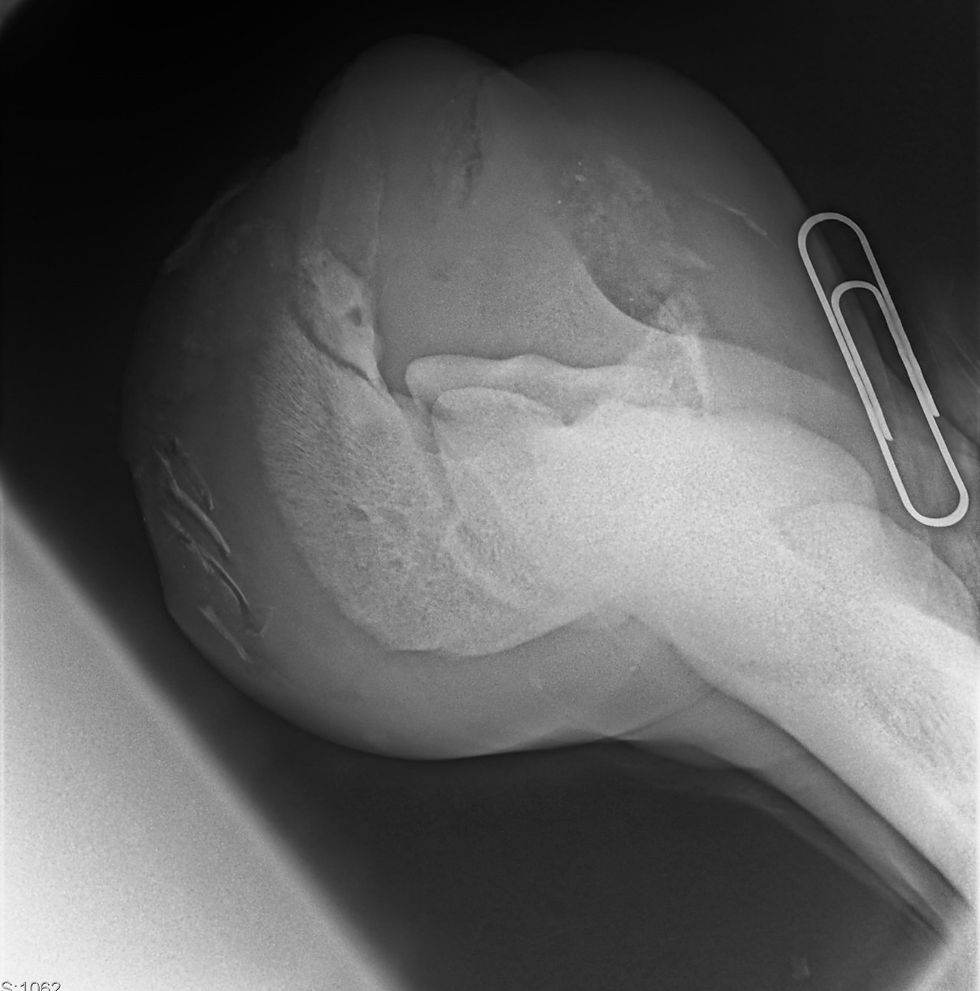
When a horse fractures its pedal bone, it will usually show signs of lameness in one leg, often quite suddenly. The horse might be unwilling to bear weight on the leg, show a strong digital pulse (a sign of inflammation), or react when hoof testers are used by a vet or farrier to check the sole. Sometimes the symptoms can look similar to other hoof problems such as an abscess or laminitis, making proper diagnosis essential. The first step is usually an X-ray, which allows the vet to see the position and type of fracture (Fig.1). However, not all fractures show up clearly on early X-rays, especially if they are hairline cracks. Sometimes a second set of X-rays is needed after a week or two when the injury becomes more visible. In some cases, advanced imaging like a CT scan or MRI is used to get a better picture (Barber et al., 2006).
Treatment
The treatment of pedal bone fractures depends on the type and severity of the break. For many fractures—especially those that are not badly displaced (meaning the bone hasn't moved much)—a non-surgical approach works well. This includes strict box rest, pain relief, and specialist shoeing. Rest gives the bone time to heal, usually over a period of 8 to 12 weeks. During this time, it’s crucial that the horse is confined to a stable and not allowed to run or play, as sudden movement could worsen the injury. Painkillers such as phenylbutazone (bute) are often prescribed in the early stages to reduce inflammation and make the horse more comfortable.
Farriery Treatment
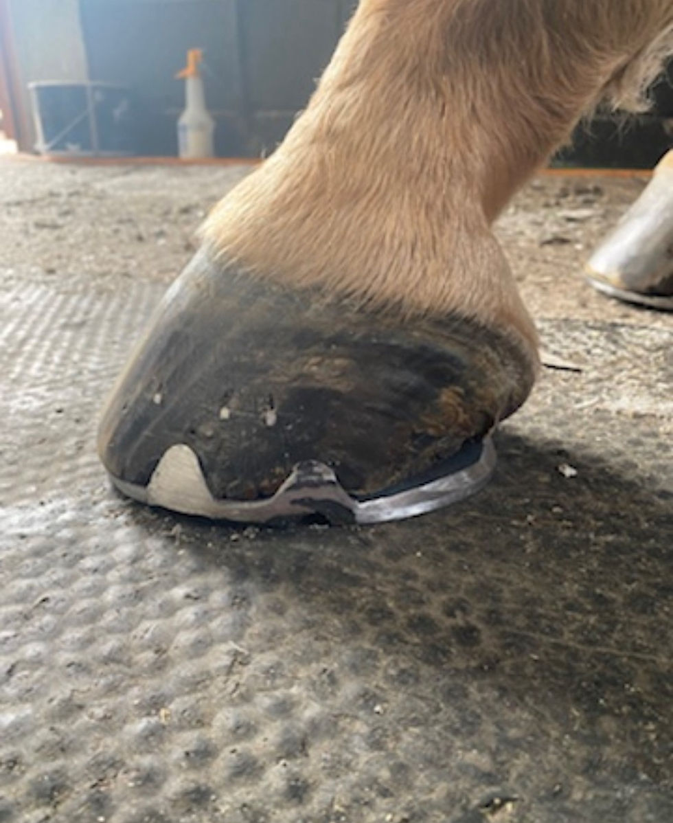
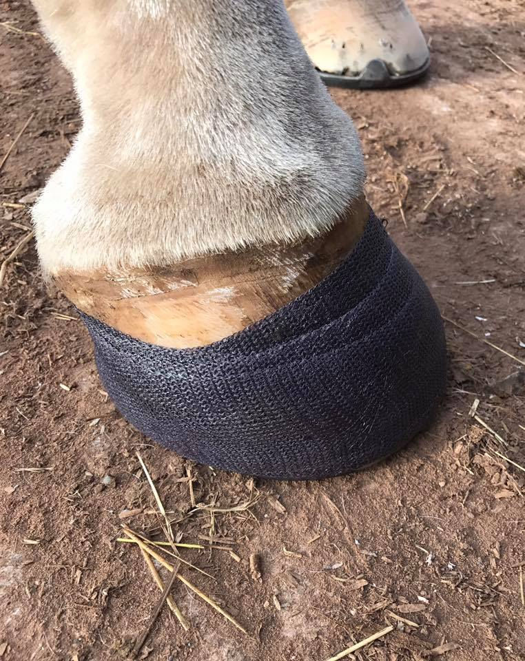
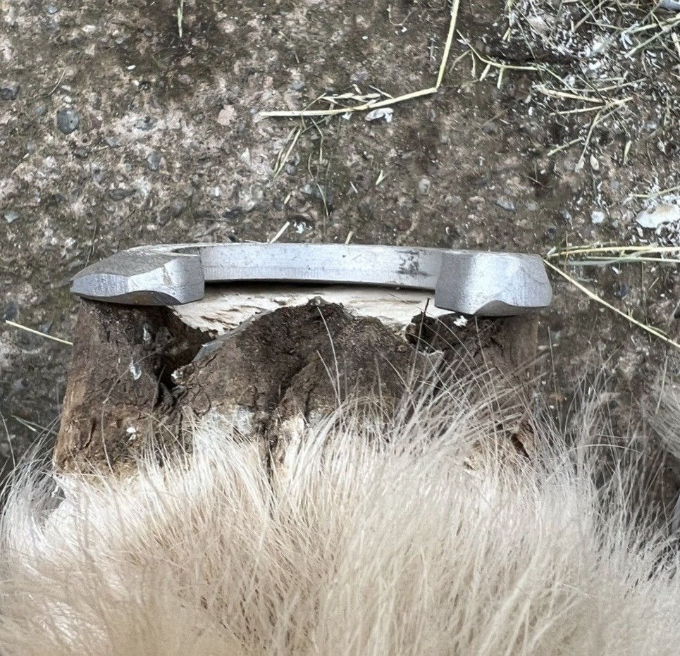
One of the most important aspects of treatment is specialist shoeing, done in collaboration with a farrier experienced in managing hoof injuries. The goal is to stabilize the hoof and reduce movement in the area where the fracture has occurred. A common solution is to fit a bar shoe, which has a solid bar connecting the two heels. This design reduces the stress on the hoof wall and helps protect the healing bone. In some cases, a soft padding material is added under the shoe to cushion the sole and reduce shock (O’Grady, 2006). Quite often a shoe with multiple clips (Fig. 2) is used to help restrict movement of the hoof capsule, a more modern approach is the use of hoof casts (Fig. 3) to aid with recovery. If the fracture is near the front of the hoof or involves the joint, the farrier may use a shoe that helps the hoof “break over” more easily, meaning it rolls forward with less strain during movement.
For more complicated fractures, especially those that involve the joint or where the bone is shattered into several pieces, surgery may be necessary. While this is rare for pedal bone injuries, there are cases where screws are used to hold the bone together, especially if the fracture runs down the centre of the bone (Richardson et al., 2003). However, operating on a pedal bone is tricky because of the risk of infection, so it is usually a last resort. Another surgical option, in cases where long-term pain remains, is neurectomy cutting the nerve that sends pain signals from the hoof. This is only considered in very specific cases where quality of life is otherwise poor, and it means the horse will no longer feel pain in that foot, which brings with it some welfare considerations.
There are also advanced treatments being researched, such as shockwave therapy which uses sound waves to stimulate healing or the use of regenerative medicine like stem cells and platelet-rich plasma (Fortier and Smith, 2008). While these techniques are promising, they are not always widely available and tend to be used in top-level performance horses or specialized clinics.
The role of the horse owner in recovery
Throughout the recovery period, the owner’s role is critical. Horses need consistent management to give them the best chance of healing. Stable rest should be enforced, and bedding should be deep and soft to reduce pressure on the hooves. Regular vet visits are needed to monitor progress, usually with follow-up X-rays every month or so. A well-balanced diet is important too, one that supports bone repair without causing the horse to become overweight from reduced exercise. The diet should contain enough calcium, phosphorus, and vitamin D, and the vet or an equine nutritionist can help make sure the balance is correct (Geor and Harris, 2013).
After the bone has healed, returning to exercise must be done gradually. Depending on the type of fracture, the horse may begin with hand walking, followed by turnout in a small paddock, and eventually light ridden work. Horses that had simpler fractures can often return to their previous level of activity, including sports like dressage, show jumping, or hacking. However, fractures involving the joint or those that were very complex may mean the horse is only suitable for light riding or pasture life afterward. In all cases, working closely with your vet and farrier will help ensure a smooth return to work.
Post recovery management
The farrier’s role doesn’t end once the fracture heals. The way the hoof is trimmed and shod after recovery is important to prevent future injuries. Poor hoof balance can place uneven stress on the pedal bone, increasing the risk of another fracture. Farriers may use X-rays to guide trimming and shoeing, ensuring that the pedal bone is in the right position within the hoof capsule (Moleman et al., 2006). Some horses may continue wearing special shoes long-term, while others may eventually return to normal shoeing or go barefoot, depending on hoof quality and workload.
Preventing pedal bone fractures is not always possible, especially in active horses, but there are steps owners can take to reduce the risk. Providing good footing in arenas and paddocks, using appropriate shoes for the horse’s activity, avoiding overworking on hard surfaces, and keeping regular farrier appointments all help reduce unnecessary strain on the hooves. In performance horses, regular check-ups with the vet and careful monitoring of any changes in movement or behaviour can help spot early signs of stress before they turn into injuries. Horses that have already had one pedal bone fracture should be managed with care, as they may be more prone to similar injuries in the future.
Conclusion
Pedal bone fractures are a serious but treatable condition. Horses with these injuries need time, careful management, and a team approach involving the vet, farrier, and owner. With prompt diagnosis, appropriate treatment, and patience during recovery, many horses go on to lead comfortable and active lives. Advances in imaging, specialist shoeing, and therapies continue to improve the outlook for these horses, and ongoing research may bring even better options in the future.
References
Barber, S. M., Witte, S. and Anderson, B. H. (2006) 'Advanced imaging of equine distal limb fractures', Veterinary Radiology & Ultrasound, 47(5), pp. 471–479.
Fortier, L. A. and Smith, R. K. W. (2008) 'Regenerative medicine for tendinous and ligamentous injuries of sport horses', The Veterinary Clinics of North America: Equine Practice, 24(1), pp. 191–201.
Geor, R. J. and Harris, P. A. (2013) Equine Applied and Clinical Nutrition. Edinburgh: Saunders Elsevier.
Moleman, M., van Heel, M. C. V., van Weeren, P. R. and Back, W. (2006) 'Hoof growth between two shoeing sessions leads to a substantial increase of the moment about the distal, but not the proximal interphalangeal joint', Equine Veterinary Journal, 38(2), pp. 170–174.
O'Grady, S. E. (2006) 'Farriery for the management of acute and chronic lameness', Veterinary Clinics of North America: Equine Practice, 22(2), pp. 303–335.
Richardson, D. W., Santschi, E. M. and Bertone, A. L. (2003) 'Fractures of the distal phalanx in horses: 25 cases (1981–1996)', Journal of the American Veterinary Medical Association, 222(11), pp. 1571–1575.
Trotter, G. W., McIlwraith, C. W., Taylor, D. S. and Segura, D. (1985) 'Management of fractures of the distal phalanx in horses: 17 cases (1976–1984)', Journal of the American Veterinary Medical Association, 187(7), pp. 721–726.


Comments