

An overview of Sidebone
- Marc Jerram

- Feb 7
- 9 min read
Introduction
Sidebone is defined as the ossification of the collateral cartilages of the distal phalanx in the equine foot and is a common finding in many horses, particularly in the forelimbs of heavier breeds and those subjected to repetitive concussion (Beasley 2024; Gray 2025). The collateral cartilages normally provide elastic support and shock absorption as the hoof deforms and expands under load, an essential function for dissipating forces transmitted through the limb during locomotion (MSD Veterinary Manual 2024). When these normally flexible cartilages become mineralised or calcified, as seen in sidebone, they lose elasticity and behave more like rigid bone, altering the biomechanics of the hoof and affecting the horse’s comfort and performance. Although many cases of sidebone are incidental and asymptomatic, understanding its development, assessment, clinical impact, diagnostic imaging, trimming requirements and shoeing strategies is crucial for farriers to support soundness and minimise long-term problems.
Prefer an audio version? Listen to the podcast at the link below:
The anatomy of the lateral cartilages underpins the pathophysiology of sidebone. Positioned on either side of the coffin bone, these paired cartilages attach proximally above the coronary band and extend distally toward the heel region. In a healthy foot, these cartilages are composed of fibrocartilaginous tissue that is relatively compliant, enabling slight deformation during stance and loading phases. This compliance facilitates hoof capsule expansion, increases circulatory dynamics, and protects deeper structures from sharp concussive forces. In sidebone, this fibrous cartilage becomes infiltrated with calcium and phosphate, progressing to bone-like tissue. Mineralisation can begin at the base of the cartilage near the junction with the distal phalanx and may extend proximally, or develop as separate centres of ossification (MSD Veterinary Manual 2024). This rigidification restricts natural hoof mechanics, reducing the capacity of the foot to absorb impact, and can thereby contribute to secondary strain on other soft tissues.
Causes
The causes and predisposing factors for sidebone are multifactorial, involving conformational, environmental, genetic and management elements. The prevailing theory is that repetitive concussion and chronic mechanical overload are primary drivers of ossification; continuous impact on unforgiving surfaces such as hard ground increases stress transmitted through the hoof to the cartilages (Wikipedia 2025; EquiMed 2025). Sidebone is particularly prevalent in heavier horses, such as draught breeds, warmbloods and cobs, likely due to their greater mass and the resultant increased axial loading of their feet (SmartPak Equine 2025; Foundation Equine 2021). Conformation faults also play a significant role by altering how forces are applied and absorbed through the limb and hoof capsule. Narrow feet with upright pasterns limit the hoof’s ability to expand and dissipate force, whereas mediolateral imbalance, toe-in or toe-out orientations and uneven limb alignment create asymmetrical loading patterns that predispose one cartilage to greater stress than the other (MSD Veterinary Manual 2024; EquiMed 2025). In such situations, sidebone may be as much a marker of longstanding mechanical imbalance and poor shock absorption as a primary causative pathology.
Static assessment of the horse
A farrier’s static assessment of a horse with suspected sidebone begins with a detailed evaluation of conformation and hoof balance while the horse stands square on a firm, level surface. The farrier should carefully observe the height and width of the heels, the alignment of the hooves relative to the limb axis, and the symmetry of the hoof capsule from medial to lateral aspects. With experience, palpation of the collateral cartilages immediately proximal to the coronary band may yield increased firmness and a lack of the usual pliability, although minor ossification may not be easily detected without imaging (MSD Veterinary Manual 2024). The static assessment also identifies hoof distortions such as flares, medial or lateral deviation and uneven growth rings, which can be indicative of past imbalance and uneven loading. It is also essential to inspect the condition of the sole and frog, as poor frog engagement and contracted heels often accompany reduced hoof expansion and may complicate sidebone progression. Static evaluation sets the stage for dynamic examination, enabling the farrier to correlate observed structural abnormalities with functional consequences.
Want to learn more about medio-lateral balance? Visit the link below:
Dynamic assessment of the horse
Dynamic assessment is equally important in revealing how the foot functions during locomotion. Observation of the horse at walk and trot on both firm and soft surfaces helps identify subtle gait abnormalities that may not be apparent during static evaluation. In horses with moderate or severe sidebone, gait changes may include shortened stride length, reluctance to bear weight on the affected limb or altered breakover patterns such as toe first or flat foot landings (Vetlexicon 2026). Watching the horse on a circle can further reveal mediolateral imbalances, with an altered loading pattern on the inside or outside limb suggesting asymmetric cartilage involvement. Dynamic assessment should not only involve surface observation but also palpation and manipulation of the distal limb to detect pain reactions or differential hoof tester responses at the heel region, although these findings are more definitive when integrated with diagnostic imaging (MSD Veterinary Manual 2024).
Clinical signs
Clinical signs associated with sidebone are variable. Many horses with radiographic evidence of collateral cartilage ossification remain sound and perform well, particularly if the condition is mild and there is no concurrent soft tissue injury (Beasley 2024; Gray 2025). Indeed, sidebone is often discovered incidentally during routine radiographic examinations or prepurchase evaluations without any reported lameness (Wikipedia 2025). When lameness does occur, it is typically mild or intermittent, exacerbated by work on hard surfaces and may coincide with localised sensitivity upon palpation. In some advanced cases, the reduced elasticity associated with ossification can contribute to secondary pathologies such as heel bruising, corns, or strain to adjacent ligaments due to altered biomechanics of the foot (EquiMed 2025). It is also recognised that fractures of extensively ossified collateral cartilages, though uncommon, can lead to acute and more severe lameness, necessitating prompt veterinary intervention.
Radiography of sidebone
The role of radiography in the diagnosis and management of sidebone cannot be overstated. Standard dorsopalmar and lateral views of the foot allow farriers and veterinarians to visualise the extent and distribution of ossification within the collateral cartilages. Radiographs not only confirm the presence of mineralised cartilage but also help determine whether the process is partial or complete, unilateral or bilateral, symmetrical or asymmetrical, which has direct implications for trimming and shoeing strategies (Vetlexicon 2026). Radiographic grading systems, often on a scale from zero (no ossification) to five (extensive ossification), provide an objective framework to monitor progression or stability over time. Additionally, specialised radiographic views such as flexed oblique projections have been shown to offer enhanced visualisation of cartilage shape, curvature abnormalities and potential fractures that might be missed on standard projections (Fenway Foundation 2015). Radiography also aids in differential diagnosis by excluding other causes of heel pain or lameness, such as pedal osteitis, distal phalanx fractures or collateral ligament injuries.

Trimming approach
Once sidebone is confirmed, whether it is symptomatic or not, a proactive trimming plan is essential to optimise hoof balance, encourage symmetrical loading and minimise excessive stress on the collateral cartilages. The overarching goal of trimming is to restore or maintain a mediolateral balanced foot with an appropriate toe length that promotes a heel first landing and effective breakover. Excessive toe length increases leverage on the distal limb segments and can exacerbate concussion forces transmitted through the collateral cartilages. Therefore, careful reduction of toe length, in concert with appropriate heel height management, encourages more efficient breakover and reduces undue leverage. Mediolateral balance is achieved by adjusting the trimming plane so that the solar surface is level and the foot bears weight evenly, which may require incremental changes over multiple trimming sessions to avoid disrupting hoof growth patterns (EquiMed 2025). Frog engagement should be maximised where possible, as this supports the digital cushion and enhances shock absorption, complementing the reduced reliance on rigid collateral cartilages.
Shoeing options
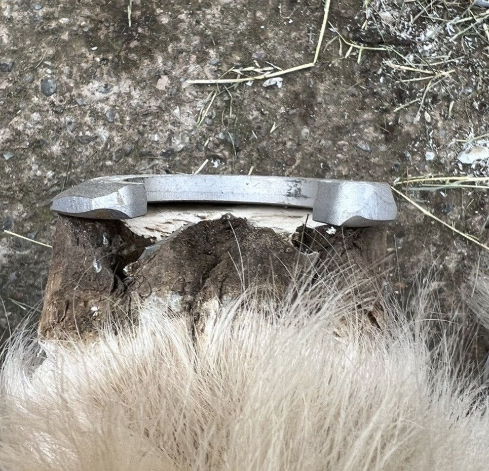
Shoeing plays a crucial role in managing sidebone by modifying how forces are transmitted through the foot. The choice of shoe should be tailored to the severity of the condition, the horse’s discipline and the degree of comfort required. A traditional sidebone shoe, which mimics the wear pattern of the previous shoe and often involves a spiral medial lift, may be used to stabilise the hoof capsule and reduce excessive movement of the ossified cartilage. This type of shoe is typically fitted full on the affected quarter to distribute load more evenly. The shoe is often made from flat bar compared to concave to reduce grab and traction which can increase strain on the ossified cartilage at the landing phase. Care must be taken to rasp down any protruding nail heads to again reduce grab as the shoe makes contact with a firm surface.
Wide web shoes can help increase ground surface area and reduce pressure on specific regions of the foot. Rolled or rockered toes may be incorporated to ease breakover and reduce lever forces during the cranial phase of the stride. In some cases, bar shoes may be beneficial by providing additional heel support and limiting excessive deformation. The use of pads, particularly supportive or shock absorbing materials, can help reduce concussion and improve comfort on hard surfaces. Pads may also promote more uniform solar loading when combined with appropriate packing materials.
The farrier must balance the need for support with the importance of allowing some degree of natural hoof movement. Overly rigid shoeing that excessively restricts expansion may exacerbate problems over time. Regular reassessment and adjustment of the shoeing plan are therefore essential (American Farriers Journal, 2004).
Shoeing strategies for horses with sidebone should be individualised based on the severity of ossification, clinical signs, workload and the horse’s overall hoof conformation. A variety of therapeutic shoeing options exist to support the foot, distribute load more evenly and limit peak pressures at the heel regions where ossified cartilage is present. In cases where sidebone is a contributor to discomfort or uneven loading, a modified shoeing approach that increases ground surface area and redistributes forces can be beneficial. For example, a full-quarter support shoe that provides additional weight bearing surface beneath the quarters may help reduce stress on a single affected collateral cartilage by promoting a broader distribution across the entire hoof (American Farriers Journal 2004). Wide web shoes can similarly increase contact area and mitigate focal pressure points.
Pads and Supportive Materials
Pads can be valuable adjuncts in the management of sidebone, particularly in horses that are sensitive to concussion. Leather, synthetic or pads made from a combination of both may be used depending on the desired level of shock absorption and support. Full sole pads can distribute load across the sole and frog, reducing peak pressures at the heels. Soft packing materials can further enhance comfort and encourage frog engagement.
The use of pads should be carefully monitored, as they can alter hoof moisture levels and obscure visual assessment of the sole. Regular removal and inspection are essential to prevent secondary issues such as thrush or sole softening, this can be reduced by using anti fungal clay or copper sulphate. When used appropriately, pads can significantly improve comfort and allow the horse to continue working within acceptable limits
It is critical to balance the need for support and protection with the preservation of natural hoof mechanics. Overly rigid shoeing that excessively restricts hoof expansion or alters normal hoof function may inadvertently contribute to further imbalance or compensatory issues. Regular reassessment of shoe fit, wear patterns and the horse’s response is essential, and adjustments should be made in collaboration with the attending veterinarian when necessary.
Prognosis
The long-term prognosis for horses with sidebone varies widely depending on the extent of ossification, presence of clinical signs, workload demands and the effectiveness of management strategies. Many horses with radiographic sidebone remain sound and fully functional in their work, particularly when sidebone is mild and discovered incidentally (Beasley 2024; Gray 2025). Indeed, radiographic findings of sidebone alone, without palpable pain or gait abnormalities, should not be considered a definitive indicator of unsoundness or reduced performance potential. As long as the horse remains comfortable under routine work, sidebone may be viewed as a structural adaptation rather than a pathological limitation.
Horses with more extensive or asymmetrical ossification that are worked predominantly on hard surfaces or at high intensity may face greater challenges. Persistent discomfort, secondary soft tissue strain or concurrent hoof abnormalities can reduce performance and comfort if not addressed. However, with appropriate trimming, thoughtful therapeutic shoeing, sensible workload management and ongoing monitoring, many affected horses maintain a good quality of life and continue to perform at acceptable levels. In cases where sidebone is associated with recurrent lameness unresponsive to conservative measures, advanced veterinary treatments including neurectomy or surgical excision of ossified cartilage may be considered, though such interventions carry a more guarded prognosis and require careful case selection (MSD Veterinary Manual 2024).
Conclusion
In conclusion, sidebone represents a complex interplay between anatomy, biomechanics, conformation, management and environmental factors. The ossification of the collateral cartilages is not inherently a career-ending condition but rather a finding that necessitates thoughtful interpretation in the clinical and functional context of the individual horse. A comprehensive assessment that integrates static and dynamic evaluation, judicious use of radiography and tailored trimming and shoeing strategies enables farriers to support soundness, comfort and performance. Ultimately, the successful management of sidebone depends on recognising the condition as part of the broader hoof function mechanism, addressing underlying imbalances, and pursuing interventions that respect the natural biomechanics of the equine foot.
References
American Farriers Journal (2004) Research Journal: May/June 2004, American Farriers Journal. Available at: https://www.americanfarriers.com/articles/7289-research-journal-may-june-2004 (Accessed: 7 February 2026).
Beasley, B. (2024) Sidebone in Horses, MSD Veterinary Manual. Merck & Co., Inc. Available at: https://www.msdvetmanual.com/musculoskeletal-system/disorders-of-the-foot-in-horses/sidebone-in-horses (Accessed: 7 February 2026).
EquiMed (2025) Sidebone, EquiMed Horse Health Matters. Available at: https://equimed.com/diseases-and-conditions/reference/sidebone (Accessed: 7 February 2026).
Fenway Foundation (2015) Diagnosing Sidebone, Fenway Foundation. Available at: https://www.fenwayfoundation.com/post/diagnosing-sidebone (Accessed: 7 February 2026).
Foundation Equine (2021) SIDEBONES, Foundation Equine. Available at: https://foundationequinenj.com/storage/app/media/FE_Client_Ed_Flyers_2021/SIDEBONESfeFlyer2021.pdf (Accessed: 7 February 2026).
Gray, L. (2025) Sidebone in Horses, SmartPak Equine. Available at: https://www.smartpakequine.com/learn-health/sidebone-horse (Accessed: 7 February 2026).
Vetlexicon (2026) Foot: lateral cartilage calcification (sidebone) in Horses, Vetlexicon Equis. Available at: https://www.vetlexicon.com/equis/surgery-orthopedic/articles/foot-lateral-cartilage-calcification-sidebone (Accessed: 7 February 2026).
Wikipedia (2025) Sidebone, Wikipedia. Available at:
https://en.wikipedia.org/wiki/Sidebone (Accessed: 7 February 2026).


Comments