

The Extensor Tendons of the Equine Forelimb and Hind Limb
- Marc Jerram

- 1 day ago
- 3 min read
Learning Outcomes
By the end of this article, you should be able to:
Describe the anatomy of the extensor tendons of the equine forelimb and hind limb, including their origin, route and insertion.
Explain the primary functions of the extensor tendons during locomotion and their role in maintaining normal limb movement.
Recognise the clinical significance of extensor tendon injuries and understand how knowledge of their anatomy assists farriers in assessing lameness and planning appropriate farriery interventions.
Definition
The extensor tendons of the equine forelimb and hind limb are strong collagenous structures that connect muscles to bone, allowing extension of the distal limb joints. They are located on the dorsal aspect of the forelimb and the cranial aspect of the hind limb and function primarily during the swing phase of the stride to extend the digit, prevent toe dragging and ensure accurate hoof placement. They also contribute to limb stability and proprioception by providing sensory feedback to the nervous system.
The main extensor tendons are the common digital extensor and lateral digital extensor in the forelimb, and the long digital extensor and lateral digital extensor in the hind limb.
Origin, Route and Insertion
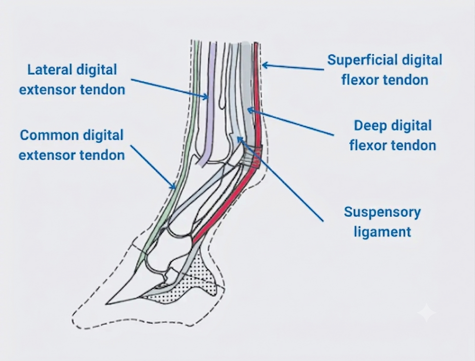
In the forelimb, the common digital extensor muscle originates from the extensor fossa on the lateral epicondyle of the humerus. Its tendon passes over the dorsal aspect of the carpus beneath the extensor retinaculum, continues down the dorsal surface of the third metacarpal bone and crosses the fetlock before inserting on the extensor process of the distal phalanx (P3). Small fibrous attachments also connect to the proximal and middle phalanges.
The lateral digital extensor muscle originates from the lateral epicondyle of the humerus, the lateral collateral ligament of the elbow and the proximal radius. Its tendon runs down the lateral aspect of the limb before joining the common digital extensor tendon in the distal metacarpus.
In the hind limb, the long digital extensor muscle originates from the extensor fossa of the femur. The tendon passes over the dorsal aspect of the hock, travels along the dorsal metatarsus and inserts on the extensor process of the distal phalanx. The lateral digital extensor originates from the lateral collateral ligament of the stifle and proximal fibula before joining the long digital extensor tendon proximal to the fetlock.
The forelimb extensor muscles are innervated by the radial nerve, while the hind limb extensors are supplied by the common fibular (peroneal) nerve.
Function
The extensor tendons extend the distal interphalangeal, proximal interphalangeal and fetlock joints during the swing phase of locomotion. This action lifts the toe clear of the ground and positions the hoof correctly before landing. Although they carry lower loads than the flexor tendons, they help maintain alignment of the digit and stabilise the dorsal aspect of the limb.
They also have an important proprioceptive role, transmitting information about limb position to the central nervous system to coordinate movement and balance. In the hind limb, the long digital extensor works with the reciprocal apparatus to produce efficient, coordinated movement between the stifle, hock and distal limb.
Clinical Relevance
Extensor tendon injuries are less common than flexor tendon injuries but are often caused by trauma because of their superficial location. Lacerations may result in toe dragging or knuckling during the swing phase, although horses usually remain weight bearing because the flexor tendons continue to support the limb.
Ultrasonography is the primary method used to assess tendon damage, while MRI may be used for more complex injuries. Treatment generally involves wound management, surgical repair when required and controlled rehabilitation. Prognosis is usually favourable because extensor tendons experience relatively low mechanical loading.
For farriers, understanding extensor tendon anatomy is important when assessing gait abnormalities, toe dragging or stumbling. Correct trimming and shoeing can improve breakover, reduce strain on injured tissues and assist rehabilitation.
Conclusion
The extensor tendons are essential for extending the digit, maintaining limb stability
and ensuring correct hoof placement during locomotion. The common and lateral digital extensor tendons of the forelimb and the long and lateral digital extensor tendons of the hind limb originate from muscles located proximally, travel along the dorsal aspect of the limb and insert on the extensor process of the distal phalanx. Although less commonly injured than the flexor tendons, their function is critical for efficient movement, making a sound understanding of their anatomy important for both farriers and veterinarians.
Learning Questions
Name the two principal extensor tendons found in the equine forelimb and the two principal extensor tendons found in the hind limb.
Where does the common digital extensor tendon insert, and why is this attachment important for normal locomotion?
What are the primary functions of the extensor tendons during the swing phase of the stride?
Which nerves supply the extensor muscles of the forelimb and hind limb, and what gait abnormality may occur if these nerves are damaged?
Why do extensor tendon injuries generally have a better prognosis than injuries to the superficial or deep digital flexor tendons?


Comments